IPPA & Beyond: Comprehensive Cardiovascular Exam Techniques Explained
In the dynamic world of medicine, mastering the art of physical diagnosis remains paramount, even amidst advanced technological marvels. Among the most critical skills a clinician can possess is the ability to perform a thorough cardiovascular examination. This systematic assessment of the heart, blood vessels, and related structures is the cornerstone for identifying normal function, subtle pathologies, and guiding subsequent management. For countless medical students and professionals, resources like the Geeky Medics Cardiovascular Exam have become indispensable, offering clear, step-by-step guidance to build confidence and competence in this essential clinical skill.
The Geeky Medics Cardiovascular Exam methodology, widely acclaimed for its practical approach, emphasizes a structured framework crucial for both learning and practical application. It transforms complex anatomical knowledge and physiological understanding into actionable examination techniques, preparing learners for everything from routine patient assessments to high-stakes Objective Structured Clinical Examinations (OSCEs).
The Enduring Importance of a Systematic Cardiovascular Assessment
While imaging technologies like echocardiography and cardiac MRI offer unparalleled insights into cardiac structure and function, the fundamental cardiovascular exam retains its irreplaceable value. Its roots trace back to early medical practices, evolving significantly with pioneers like Sir James Mackenzie in the 19th century, who championed a systematic approach to patient assessment. Today, it remains a critical first line of defense in diagnosing a spectrum of conditions, from common ailments to life-threatening emergencies.
A meticulous cardiovascular examination can reveal vital clues about conditions such as hypertension, various forms of heart failure, coronary artery disease, and diverse valvular diseases. By identifying subtle signs early, clinicians can significantly reduce misdiagnosis rates and initiate timely interventions, aligning with recommendations from leading organizations like the American Heart Association. Beyond its diagnostic power, the exam hones crucial clinical skills, including patient interaction, empathetic communication, and the synthesis of findings into coherent clinical reasoning. It's a testament to the power of observation and skilled touch in medical practice.
Deconstructing the IPPA Framework: Core Techniques Explained
The cardiovascular exam is traditionally taught using the IPPA framework: Inspection, Palpation, Percussion, and Auscultation. Each component serves a distinct purpose, building a comprehensive picture of the patient's cardiovascular health.
1. Inspection: The Art of Observation
Inspection begins the moment you meet the patient and continues throughout the entire examination. It involves a systematic visual assessment, looking for tell-tale signs that betray underlying cardiovascular issues.
- General Appearance: Observe the patient's posture, comfort level, and signs of distress (e.g., shortness of breath, pallor). Is there evidence of cachexia (severe weight loss), which can be associated with chronic heart failure?
- Hands: Look for finger clubbing (cyanotic heart disease, infective endocarditis), Janeway lesions (painless, red spots on palms/soles, infective endocarditis), Osler's nodes (tender, red lesions on finger/toe pads, infective endocarditis), splinter hemorrhages (nail bed streaks, endocarditis), and xanthomata (fatty deposits, hyperlipidemia).
- Arms: Check for evidence of previous procedures (e.g., scars from angiograms, AV fistulas).
- Face: Assess for conjunctival pallor (anemia), malar flush (mitral stenosis), corneal arcus (lipid deposits, hyperlipidemia), and xanthelasma (fatty deposits around eyes).
- Neck: Crucially, assess for Jugular Venous Distension (JVD), which indicates elevated right atrial pressure, often seen in right-sided heart failure. Also, observe for carotid pulsations and any visible thrills.
- Chest and Precordium: Look for chest deformities (e.g., pectus excavatum or carinatum), scars from previous cardiac surgeries (e.g., sternotomy for CABG or valve replacement), and any visible apical impulse or abnormal pulsations (heaves).
- Lower Legs & Feet: Inspect for peripheral edema (bilateral often suggests heart failure; unilateral suggests DVT), skin changes, and signs of peripheral vascular disease (e.g., hair loss, trophic changes, ulcers).
2. Palpation: The Touch for Truth
Palpation involves using your hands and fingertips to feel for specific characteristics and abnormalities.
- Pulses: Systematically palpate various peripheral pulses for rate, rhythm, volume, and character. This includes radial, brachial, carotid, femoral, popliteal, dorsalis pedis, and posterior tibial pulses. Abnormalities like a "waterhammer" pulse (aortic regurgitation) or pulsus paradoxus (cardiac tamponade) provide crucial diagnostic clues.
- Precordial Palpation:
- Apex Beat (Point of Maximal Impulse - PMI): Locate the apex beat, usually found in the 5th intercostal space, midclavicular line. Note its location, size, and character. Displacement laterally or inferiorly can indicate cardiomegaly.
- Thrills: Palpate across the precordium for thrills, which are palpable vibrations caused by turbulent blood flow, indicative of significant murmurs.
- Heaves: Feel for sustained, outward thrusts during systole, known as heaves or lifts, often signifying ventricular hypertrophy (e.g., parasternal heave in right ventricular hypertrophy).
3. Percussion: A Fading but Foundational Technique
While less commonly used today due to the widespread availability of imaging, percussion historically involved tapping the chest to estimate heart size and borders. A dull sound would indicate the presence of cardiac tissue or fluid (e.g., pericardial effusion). Modern imaging, particularly echocardiography, offers far more precise measurements, but understanding its historical role provides context.
4. Auscultation: Listening to the Heart's Song
Auscultation, performed with a stethoscope, is arguably the most critical component. It involves listening attentively to the heart sounds, murmurs, and any extra sounds. Always auscultate in a quiet environment with the patient adequately exposed.
- Key Auscultatory Areas:
- Aortic Area: Right 2nd intercostal space, sternal border.
- Pulmonary Area: Left 2nd intercostal space, sternal border.
- Erb's Point: Left 3rd intercostal space, sternal border (useful for hearing both aortic and pulmonary sounds).
- Tricuspid Area: Left 4th/5th intercostal space, sternal border.
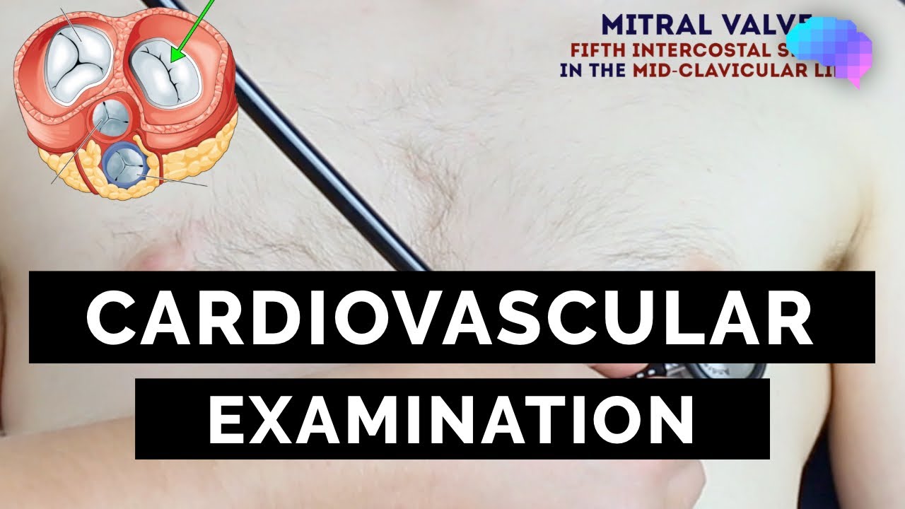
- Mitral (Apical) Area: Left 5th intercostal space, midclavicular line.
- Heart Sounds (S1 and S2):
- S1 ("lub"): Represents the closure of the atrioventricular (mitral and tricuspid) valves, best heard at the apex.
- S2 ("dub"): Represents the closure of the semilunar (aortic and pulmonary) valves, best heard at the base (aortic and pulmonary areas). Physiological splitting of S2 (where the aortic and pulmonary components separate during inspiration) is normal. Pathological splitting (wide, fixed, or paradoxical) indicates underlying issues.
- Extra Sounds: Listen for S3 (early diastolic gallop, often associated with heart failure), S4 (late diastolic gallop, often associated with stiff ventricle), pericardial friction rubs (pericarditis), and clicks (prosthetic valves).
- Murmurs: These are turbulent blood flow sounds. Characterize them by:
- Timing: Systolic (between S1 and S2) or Diastolic (between S2 and S1).
- Location: Where they are best heard.
- Radiation: To the neck (aortic stenosis) or axilla (mitral regurgitation).
- Quality: Blowing, harsh, rumbling, musical.
- Intensity: Graded from I/VI (faint) to VI/VI (audible without stethoscope).
- Pitch: High or low.
- Maneuvers: How they change with respiration, position changes, or Valsalva maneuver.
A Systematic Approach: Step-by-Step for Clinical Mastery
Performing a comprehensive cardiovascular exam requires a systematic workflow to ensure no vital signs are missed. Here’s a detailed, step-by-step guide, mirroring the structured approach championed by Geeky Medics:
- Preparation & Introduction:
- Hand Hygiene & PPE: Always begin by washing your hands and don appropriate personal protective equipment if necessary.
- Introduction: Introduce yourself clearly, stating your name and role.
- Patient Confirmation: Verify the patient's name and date of birth.
- Explanation & Consent: Briefly explain the examination in patient-friendly language and obtain explicit consent to proceed.
- Positioning & Exposure: Adjust the head of the bed to a 45° angle. Ensure adequate exposure of the patient's chest (from the waist up), offering a blanket for modesty. Exposure of the lower legs is also often helpful.
- History Taking (Brief): Before touching the patient, gather essential history. Ask about presenting symptoms (chest pain, dyspnea, palpitations, syncope, edema), relevant medical history (hypertension, diabetes, previous MIs), risk factors (smoking, family history), and medications.
- General Inspection: Observe the patient's overall appearance, comfort, and any visible signs from a distance.
- Hands & Arms: Proceed with detailed inspection and palpation as described above. Measure blood pressure in both arms.
- Face & Neck: Inspect the face, then assess the jugular venous pressure (JVP) and palpate (one at a time) and auscultate the carotid arteries for bruits.
- Chest (Precordial Exam):
- Inspection: Look for deformities, scars, visible pulsations.
- Palpation: Palpate the apex beat, thrills, and heaves.
- Auscultation: Systematically auscultate all five areas using both the diaphragm and bell of your stethoscope. Pay close attention to S1, S2, murmurs, and extra sounds. Listen over the carotids as well.
- Abdomen: Briefly inspect and palpate for abdominal aortic pulsation or distension (ascites).
- Lower Legs & Feet: Inspect for edema, skin changes, and palpate peripheral pulses (femoral, popliteal, dorsalis pedis, posterior tibial).
- Completion: Thank the patient, help them re-cover, and wash your hands. Summarize your findings and suggest further investigations if necessary.
Beyond the Basics: Advanced Insights & Common Pitfalls
While the IPPA framework provides a robust foundation, true mastery of the cardiovascular exam lies in developing clinical reasoning and avoiding common pitfalls. Don't just perform the steps; interpret the findings in context. For instance, a displaced apex beat in a patient with a history of hypertension strongly suggests left ventricular hypertrophy, while a new murmur post-MI could indicate a ruptured papillary muscle.
Common Pitfalls to Avoid:
- Inadequate Exposure: Trying to auscultate through clothing can obscure vital sounds.
- Rushing: A hurried exam often misses subtle but significant findings. Allow enough time for quiet auscultation.
- Ignoring Patient Comfort: Failing to explain steps or provide privacy can lead to a less cooperative patient and a less effective exam.
- Not Correlating Findings: An isolated finding is less useful than synthesizing all signs with the patient's history.
- Misinterpreting Sounds: Distinguishing between physiological and pathological sounds takes practice. Listen to many hearts!
Always consider the "whole patient." A comprehensive understanding of anatomy, physiology, and pathology will elevate your examination from a rote procedure to a powerful diagnostic tool. If findings are unclear or suggest significant pathology, further investigations like an ECG, chest X-ray, or echocardiogram are warranted, and specialist consultation may be necessary.
Conclusion
The cardiovascular exam, while an ancient practice, remains a cornerstone of modern medicine. Its systematic application, as meticulously taught by resources like the Geeky Medics Cardiovascular Exam, empowers clinicians to accurately assess heart function, identify abnormalities, and ultimately provide better patient care. By diligently practicing inspection, palpation, percussion, and auscultation within a structured framework, medical students and seasoned professionals alike can refine their clinical skills, ensuring that no subtle clue is overlooked in the journey toward accurate diagnosis and effective treatment. Embrace the art of the physical exam; your patients depend on it.